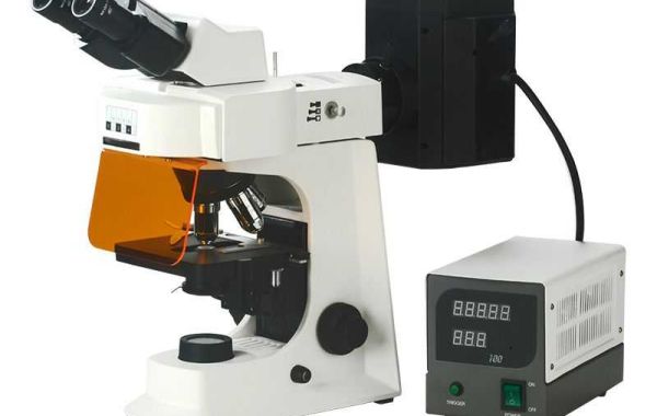
Here's why knowing how to prepare samples for a fluorescence microscope is crucial:
- High-Quality Images: Proper sample preparation is essential for obtaining high-quality images with a fluorescence microscope. The preparation steps like fixation, staining, and mounting can significantly impact the final image. Proper techniques ensure the target structures are well-preserved, fluorescent labels bind effectively, and background noise is minimized. This all leads to clear and informative images for your analysis.
- Specificity and Accuracy: Fluorescence microscope relies on specific labeling of target molecules with fluorophores. Knowing proper sample preparation techniques helps ensure these labels bind only to the structures you're interested in. This specificity minimizes background fluorescence from unwanted structures and allows for accurate identification and analysis of your target molecules.
- Preserving Cellular Structures: Many fluorescence microscopy experiments involve studying cellular components. Sample preparation techniques like fixation help preserve the morphology (shape and organization) of these structures. Without proper fixation, cells can degrade or collapse, making it difficult to interpret the results and draw meaningful conclusions.
- Fluorescence Intensity:The intensity of the fluorescent signal is directly related to the number of fluorophores bound to the target molecule. Proper sample preparation techniques optimize binding efficiency, leading to brighter fluorescence and a stronger signal. This allows for better detection and analysis of even low-abundance molecules within the cell.
- Optimizing Experiments:Knowing various sample preparation methods allows you to choose the technique best suited for your specific experiment. For example, if you're studying live cells, you might use a different approach compared to studying fixed tissues. Understanding these options allows you to optimize your experiment for the desired information.
- Minimizing Errors:Improper sample preparation can introduce artifacts or errors into your data. For instance, inadequate fixation can lead to loss of structures or non-specific staining. By understanding proper techniques, you can minimize these errors and ensure the validity of your results.
In conclusion, knowing how to prepare samples for fluorescence microscope is essential for obtaining high-quality, accurate, and informative data from your experiments. It allows you to visualize cellular structures clearly, achieve specific labeling, and optimize your overall analysis.


