What are Dyed Polystyrene Latex Particles?
Polystyrene particles (PSP) are commercially available in different sizes, ranging from 15 nanometers to several micrometers, with narrow size distributions and various surface chemistries. Additionally, polystyrene is generally considered inert and nontoxic, making it safe for use in a variety of biological assays. Dyed polystyrene latex particles or colored latex beads have excellent multiplexing capability and have the following characteristics: a. Diversity, that is, diversity of particle size, color, and color depth; b. High labeling efficiency; c. Stability, it has good chemical and physical properties in aqueous solution. These characteristics provide a strong guarantee for the rapid development and application of colored latex particles in medical diagnostic technology.
Shop for Colored Polystyrene Particles
| Black Polystyrene Particles | Red Polystyrene Particles |
| Blue Polystyrene Particles | Violet Polystyrene Particles |
| Green Polystyrene Particles | Yellow Polystyrene Particles |
| Orange Polystyrene Particles |
At present, there are three main methods for preparing colored latex particles: the first is to copolymerize dye monomers and latex monomers; the second is to prepare latex particles first, then modify them with active groups on the surface of the particles, and finally combine the dye molecules by covalent cross-linking to the particle surface; a third method is to prepare colored latex particles by physically trapping or absorbing dye molecules.
Immunochromatographic Assay: Leveraging Colored Polystyrene Particles
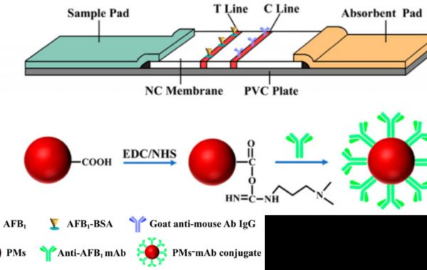
Immunochromatographic assay (ICA), also known as lateral flow assay (LFA), is a rapid analysis technology with simplicity, speed, and sensitivity advantages. ICA is one of the most successful and widely used point-of-care tests (POCT) and has been successfully used for human chorionic gonadotropin (hCG) detection, serodiagnostic assays, cancer detection, cardiac markers and infectious microorganism detection. ICA has been widely used in many fields such as medicine and food safety. To enhance the sensitivity of colorimetric ICA, labeled nanomaterials need to exhibit deep and bright color signals. Utilizing new colored nanomaterials as labeled probes is an important strategy to improve sensitivity. Wang et al. developed crimson red 0.2000 µm polystyrene microspheres (PMs)-based ICA for sensitive and quantitative detection of AFB1 in corn. PMs-ICA is more sensitive than CG (colloidal gold)-ICA, and results can be obtained with the naked eye rather than using a reader. Nagaoka et al developed a new visual immunodiagnostic method using red latex beads to detect urinary IgG4. Taking those with ICT antigen test positive as the standard (136/156), the sensitivity is 87.2%. Comparing gold nanoparticles with latex beads, it can be observed that lateral flow assays using latex beads on the conjugation pad have higher sensitivity and specificity than assays using gold nanoparticles.
Latex Agglutination Assays: The Role of Dyed Polystyrene Particles
Latex agglutination is the change in agglutination capacity observed when a sample containing a specific antigen is mixed with antibodies coated on the surface of latex particles. Agglutination can also be reversed by coating latex particles with specific antigens and testing the sample for the presence of specific antibodies. Latex agglutination assays have the advantage of rapid results, often determined within minutes, and this type of reaction does not require expensive equipment to evaluate the sample. A user-friendly latex agglutination assay was developed and evaluated for the serodiagnosis of human brucellosis. The assay was obtained by coating blue latex beads with Brucella lipopolysaccharide and drying the activated beads onto a white agglutination card. For initial serum samples collected from patients with culture-confirmed brucellosis, the assay had a sensitivity of 89.1% (95% CI, 76-96) and a specificity of 98.2% (95% CI, 96-99). Grace et al. developed a rapid diagnostic test for porcine JEV diagnosis and serosurveillance using violet-dyed polystyrene beads (0.80 μm) that can be easily applied in the field and is useful to fill the gap between existing diagnostic methods and surveillance programs.
Advantages of Colored Polystyrene Particles in Immunoassays
Latex particles formed from amorphous polymer (usually polystyrene) represent a class of particles that have applications in many different aspects of biotechnology. These monodisperse particles range in size from 20 nm to 160 µm, with 100-400 nm covering the typical range used in lateral flow immunoassays. Colored polystyrene beads are inexpensive. Second, because they are doped with organic dyes, they are available in a variety of colors, with red, blue, black, and green being the most popular choices for lateral flow assays. The versatility of latex particles in color selection and compatibility with many biological matrices make them ideal candidates for labeling in many lateral flow immunoassays.
References
- Nagaoka, F., Itoh, M., Samad, M. S., Takagi, H., Weerasooriya, M. V., Yahathugoda, T. C., … Kimura, E. (2013). Visual detection of filaria-specific IgG4 in urine using red-colored high density latex beads. Parasitology international, 62(1), 32-35.
- Wang, J., Li, X., Shen, X., Zhang, A., Liu, J., Lei, H. (2021). Polystyrene microsphere-based immunochromatographic assay for detection of aflatoxin B1 in maize. Biosensors, 11(6), 200.
- Abdoel, T. H., Smits, H. L. (2007). Rapid latex agglutination test for the serodiagnosis of human brucellosis. Diagnostic microbiology and infectious disease, 57(2), 123-128.
- Grace, M. R., Dhanze, H., Pantwane, P., Sivakumar, M., Gulati, B. R., Kumar, A. (2019). Latex agglutination test for rapid on-site serodiagnosis of Japanese encephalitis in pigs using recombinant NS1 antigen. Journal of Vector Borne Diseases, 56(2), 105-110.


